Diabetic Foot Examination: A Comprehensive Guide
Annual foot inspections, including vascular, neurological, and skin assessments, are crucial for diabetic patients, as detailed in clinical guidelines and reports.
Diabetic foot exams are a cornerstone of preventative care for individuals living with diabetes. These comprehensive evaluations aim to identify risk factors and early signs of complications, ultimately reducing the likelihood of foot ulcers, infections, and potential amputations.
Regular examinations, ideally performed annually by a healthcare professional trained in foot care – physicians (MD, DO, DPM) or advanced practice providers – are vital. The exam encompasses a detailed assessment of vascular function, neurological integrity, and skin health.
Registered Nurses (RNs) play a key role in increasing exam rates through toolkits, providing patient education on proper foot care, and documenting findings within Electronic Health Records (EHRs).
Why are Diabetic Foot Exams Important?
Diabetic foot exams are critically important due to the increased risk of lower-extremity complications in individuals with diabetes. Neuropathy, diminished sensation, and peripheral arterial disease (PAD) can lead to unnoticed injuries and impaired healing.
Early detection of these issues through regular exams allows for timely intervention, preventing minor problems from escalating into serious infections or ulcers. Comprehensive assessments, including vascular pulse palpation, are essential for defining overall risk status.
Proactive foot care, guided by exam findings, significantly reduces the potential for amputation and improves patients’ quality of life.
Risk Factors for Diabetic Foot Complications
Several factors elevate the risk of diabetic foot complications. Peripheral neuropathy, a common diabetes complication, diminishes protective sensation, increasing injury risk. Peripheral arterial disease (PAD) impairs blood flow, hindering wound healing.
Poorly controlled blood sugar levels exacerbate both neuropathy and PAD. Foot deformities, like bunions or hammertoes, create pressure points prone to ulceration. A history of previous foot ulcers significantly increases recurrence risk.
Inadequate footwear and improper foot care practices also contribute. Smoking further compromises vascular health, compounding the risk. Recognizing these factors is vital for targeted prevention.
Components of a Diabetic Foot Examination
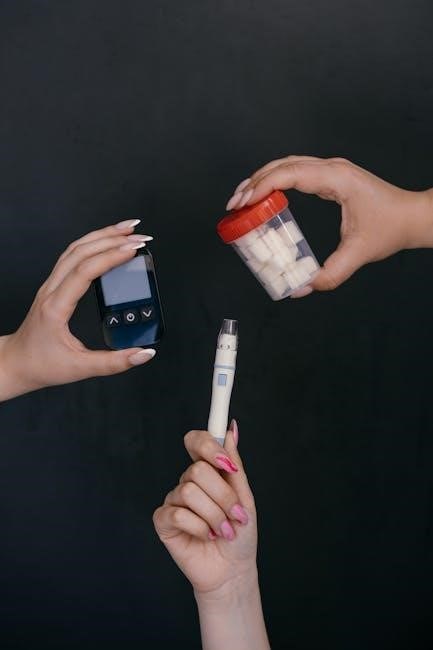
A thorough exam includes patient history, visual inspection, vascular assessment (pulses), neurological testing (monofilament, pinprick), and musculoskeletal/footwear evaluation.
Patient History
Gathering a detailed patient history is the foundational step in a comprehensive diabetic foot exam. This involves a focused medical history, specifically exploring conditions impacting foot health, such as peripheral vascular disease, neuropathy, and prior diabetes-related complications.
Crucially, document any foot ulcer history – location, size, healing time, and prior treatments. A complete medication review is also essential, noting any drugs that could affect circulation, nerve function, or wound healing.
Understanding the patient’s lifestyle, including footwear choices and daily activity levels, provides valuable context. This holistic approach allows for personalized risk assessment and targeted interventions.
Medical History Relevant to Foot Health
A thorough medical history must prioritize conditions impacting foot health. Specifically, inquire about the duration of diabetes, glycemic control (HbA1c levels), and history of peripheral vascular disease (PVD).
Neuropathy, both peripheral and autonomic, is critical to assess, noting symptoms like numbness, tingling, or burning pain. Previous hospitalizations related to foot issues, like infections or ulcers, require detailed documentation.
Furthermore, explore a history of smoking, hypertension, hyperlipidemia, and renal disease, as these contribute to vascular complications. Understanding these factors informs risk stratification and guides examination priorities.
Foot Ulcer History
Detailed documentation of any prior foot ulcers is paramount. Ascertain the location, size, depth, and duration of previous ulcers. Investigate whether prior ulcers healed completely, partially, or required surgical intervention, such as amputation.
Crucially, determine the cause of previous ulceration – was it related to trauma, pressure, infection, or vascular insufficiency? Explore prior treatments received, including antibiotics, offloading devices, and wound care regimens.
Recurrence of ulcers signifies heightened risk; understanding the pattern of previous ulcerations helps predict future vulnerability and tailor preventative strategies accordingly.
Medication Review
A thorough medication review is essential, focusing on drugs impacting peripheral circulation or wound healing. Specifically, assess for the use of anticoagulants, antiplatelet agents, and medications known to cause peripheral vasoconstriction.
Review the patient’s diabetes management regimen, including oral hypoglycemic agents and insulin therapy, to evaluate glycemic control. Corticosteroids, even topical applications, can impair wound healing and should be noted.
Identify any medications potentially contributing to edema or neuropathy. Document all medications, dosages, and duration of use to comprehensively understand potential influences on foot health.
Visual Inspection
A detailed visual inspection is paramount, beginning with a comprehensive assessment of skin integrity. Look for any breaks in the skin, ulcerations, blisters, or areas of redness, swelling, or warmth. Carefully examine the interdigital spaces for signs of fungal infection or maceration.
Identify any foot deformities, such as hammertoes, claw toes, bunions, or Charcot foot, as these can predispose to pressure points and ulcer development. Note the presence of calluses or corns, indicating areas of repetitive friction.
Assess for signs of infection, including purulent drainage, cellulitis, or osteomyelitis.
Skin Integrity Assessment
Meticulous skin assessment is fundamental, involving a thorough examination for any disruptions. Specifically, identify breaks in the skin, open sores, blisters, or areas exhibiting redness, swelling, or unusual warmth. Pay close attention to the interdigital spaces, checking for maceration or fungal infections.
Evaluate for dryness, cracking, or scaling, as these can increase vulnerability to infection. Document any existing calluses or corns, noting their location and size. Assess the skin’s temperature bilaterally, looking for discrepancies that might suggest inflammation or infection.
Deformity Identification
Careful observation for foot deformities is essential during the examination. Common deformities include hammertoes, claw toes, bunions, and Charcot foot – a severe condition causing bone and joint destruction. Palpate the bony prominences and assess for any unusual swelling or instability;
Evaluate the alignment of the toes and the overall foot structure. Note any areas of pressure or friction caused by deformities, as these are prone to ulceration. Document the severity of each deformity and its potential impact on gait and footwear choices. Early identification allows for preventative measures.
Looking for Signs of Infection
Diligent assessment for infection is paramount, as diabetic foot ulcers can rapidly progress. Examine the skin for redness, warmth, swelling, and any visible drainage. Note the color and consistency of any discharge – purulent exudate suggests infection.
Assess for local signs of inflammation and systemic symptoms like fever or chills. Palpate surrounding tissues for tenderness and crepitus. Document any breaks in the skin, even minor ones, as entry points for bacteria. Prompt recognition and treatment are vital to prevent serious complications like osteomyelitis.
Vascular Examination
A thorough vascular assessment is critical for identifying Peripheral Arterial Disease (PAD), a significant risk factor for diabetic foot complications. This begins with palpation of the posterior tibial and dorsalis pedis pulses, characterizing them as present or absent.
Assess the overall lower-extremity risk status, as PAD significantly impacts healing potential. Evaluate skin temperature and color; coolness or pallor can indicate reduced blood flow. Look for signs of claudication – pain with exercise relieved by rest. Accurate vascular assessment guides appropriate interventions and preventative care.
Palpation of Posterior Tibial Pulse
Locate the posterior tibial pulse by palpating behind the medial malleolus, the bony prominence on the inner side of the ankle; Use gentle, circular pressure with your fingertips. Assess the pulse’s quality – is it bounding, strong, weak, or absent?
Document your findings clearly. An absent or diminished pulse suggests potential peripheral arterial disease. Compare the pulse strength to the contralateral foot. Consistent and accurate palpation is essential for identifying vascular compromise, a key component of diabetic foot risk assessment and guiding further diagnostic testing.
Palpation of Dorsalis Pedis Pulse
Identify the dorsalis pedis pulse by locating it on the top of the foot, between the first and second metatarsal bones. Apply gentle, circular pressure with your fingertips to assess pulse presence and quality – bounding, strong, weak, or absent.
Accurate documentation is vital. A diminished or absent pulse raises concerns about peripheral arterial disease. Always compare the pulse strength bilaterally. Consistent assessment of the dorsalis pedis pulse, alongside the posterior tibial pulse, provides a comprehensive vascular evaluation, crucial for managing diabetic foot risk.
Assessment of Peripheral Arterial Disease (PAD)
Evaluating for PAD is essential, as it significantly impacts lower-extremity risk in diabetic patients. This assessment builds upon pulse palpation – specifically, the presence or absence of the posterior tibial and dorsalis pedis pulses. Diminished or absent pulses strongly suggest potential arterial insufficiency.
Further investigation may be needed if PAD is suspected. Comprehensive assessment defines overall risk status. Early detection allows for timely intervention, potentially preventing ulceration and amputation. Accurate PAD assessment is a cornerstone of effective diabetic foot care.
Neurological Examination
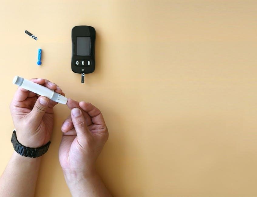
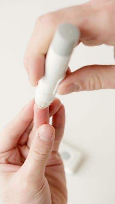
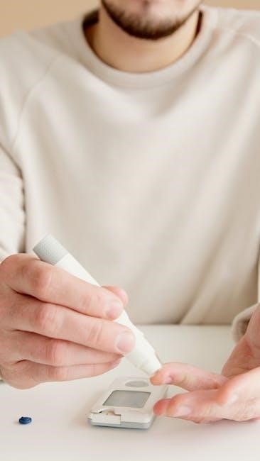
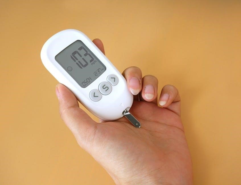
A thorough neurological exam is paramount in diabetic foot care, as neuropathy is a major contributor to foot complications. This involves assessing sensory function using tools like the 10-gram monofilament test, determining a patient’s ability to perceive light touch. Pinprick sensation testing further evaluates pain perception, identifying areas of diminished or absent feeling.
Vibration perception testing adds another layer of assessment. These tests help identify peripheral neuropathy early, enabling proactive interventions and patient education to prevent ulceration and limb loss. Consistent neurological evaluation is vital.
Monofilament Testing
Monofilament testing is a cornerstone of the neurological examination for diabetic patients, serving as a simple yet effective method to assess protective sensation. A 10-gram monofilament is applied to specific locations on the foot – typically the first, third, and fifth metatarsal heads – to determine if the patient can detect the pressure.
Inability to feel the monofilament indicates a loss of protective sensation, significantly increasing the risk of foot ulceration. This test helps identify patients needing heightened foot care education and more frequent monitoring, preventing potentially serious complications.
Pinprick Sensation Testing
Pinprick sensation testing complements monofilament testing in evaluating a patient’s neurological function and risk for diabetic foot complications. Utilizing a sterile pin or needle, the examiner gently pricks designated areas of the foot – often the same locations as monofilament testing – to assess the patient’s ability to perceive sharp stimuli.
A diminished or absent response suggests neuropathy and impaired protective sensation. While monofilament testing assesses pressure, pinprick testing evaluates pain perception, providing a more comprehensive neurological profile. This combined approach aids in identifying individuals requiring intensive foot care interventions.
Vibration Perception Testing
Vibration perception testing is a valuable component of a comprehensive diabetic foot examination, assessing the function of the peripheral nerves. A vibrating tuning fork (typically 128 Hz) is gently placed on bony prominences of the foot, such as the malleoli or the distal phalanx of the great toe.
The patient is asked to indicate when they feel the vibration stop. Decreased or absent vibration sensation indicates neuropathy, signifying nerve damage and an increased risk of foot ulceration. This test helps identify early nerve dysfunction, even before changes are detectable with monofilament or pinprick testing.
Musculoskeletal Examination
Musculoskeletal assessment is a critical part of the diabetic foot exam, evaluating foot structure and biomechanics to identify potential problems. This involves assessing the range of motion in the foot and ankle joints, looking for limitations or deformities.
The examiner should observe the foot’s arch height, noting any flattening or excessive curvature. Palpation can reveal bony prominences or areas of tenderness. Assessing gait and weight distribution provides insight into biomechanical stresses. Identifying these factors helps predict ulcer risk and guide appropriate interventions, like orthotics.
Range of Motion Assessment
Range of motion (ROM) assessment is a key component, evaluating flexibility and movement within the foot and ankle joints. Both active and passive ROM should be assessed, noting any limitations or pain during movement.
Specifically, dorsiflexion, plantarflexion, inversion, and eversion are evaluated. Limited ROM can alter gait mechanics and increase pressure points, elevating ulcer risk. Stiffness may indicate joint damage or neuropathy-related changes. Documenting any restrictions or abnormalities is crucial for comprehensive care planning and monitoring disease progression.
Foot Structure and Biomechanics
Assessing foot structure and biomechanics involves evaluating arch height, alignment, and weight distribution. Look for deformities like hammertoes, claw toes, or bunions, which can create areas of high pressure.
Observe the patient’s gait to identify abnormal patterns contributing to stress. Neuropathy can cause altered biomechanics and foot deformities. Proper assessment helps identify risk factors for ulcer development; Document any structural abnormalities or gait deviations, as these findings guide interventions like orthotics or footwear modifications to optimize foot health.
Footwear Assessment

Footwear plays a critical role in preventing complications. Assess shoes for proper fit – adequate depth and width to accommodate any deformities, and sufficient toe box space. Examine the interior for seams or objects that could cause pressure or irritation.
Evaluate sock material; seamless, moisture-wicking socks are preferred. Educate patients on the importance of well-fitting footwear and avoiding walking barefoot. Ill-fitting shoes contribute significantly to ulceration. Document footwear type, fit, and sock condition as part of the comprehensive exam.
Proper Shoe Fit
Ensuring correct shoe fit is paramount for diabetic patients. Shoes should accommodate foot deformities without causing pressure points. A thumb’s width of space should exist between the longest toe and the shoe’s end. Width must be appropriate, avoiding constriction.
Assess heel fit – minimal slippage is ideal. Patients should be instructed to shop for shoes later in the day when feet are most swollen. Regularly check shoes for internal irregularities. Proper fit minimizes friction and shear forces, reducing ulcer risk. Document fit assessment findings meticulously.
Sock Evaluation
Appropriate sock selection is a vital, often overlooked, component of diabetic foot care. Seams should be minimal or absent to prevent irritation and pressure. Materials like cotton are discouraged due to moisture retention; synthetic or wool blends are preferred for wicking properties.
Socks should not be constricting, hindering circulation. Evaluate for proper size – avoiding bunching or tightness. Patients should change socks daily, or more frequently if moisture is present. Educate on the importance of avoiding tight elastic bands. Document sock type and condition during each examination.
Documentation and Reporting
Detailed records, utilizing pre-populated EHR templates, are essential for tracking exam findings, coding, and billing for comprehensive diabetic foot evaluations.
Electronic Health Record (EHR) Templates
Utilizing standardized EHR templates is paramount for efficient and thorough documentation of diabetic foot examinations. These templates should facilitate consistent recording of key findings, including vascular assessment results – specifically, the presence or absence of posterior tibial and dorsalis pedis pulses.
Furthermore, templates must incorporate neurological examination data, such as monofilament testing and pinprick sensation results, alongside detailed observations regarding skin integrity and any identified foot deformities.
Pre-populated fields streamline the process, reducing documentation time for healthcare professionals, while ensuring all critical elements are captured. Proper template use supports accurate coding and billing practices, and enhances continuity of care through readily accessible patient information.
Coding and Billing for Diabetic Foot Exams
Accurate coding and billing are essential for diabetic foot examinations, reflecting the comprehensive nature of the assessment. Proper documentation, utilizing standardized EHR templates with detailed findings – including vascular pulse palpation and neurological testing results – is crucial for justifying billed services.
Healthcare providers must adhere to current coding guidelines, ensuring appropriate assignment of CPT codes based on the examination’s complexity and components performed.
Thorough documentation supports medical necessity and minimizes claim denials, while maximizing appropriate reimbursement for these vital preventative care services. Staying updated on coding changes is paramount for compliant billing practices.
Patient Education
Educating patients on proper foot care, footwear choices, and recognizing infection signs is vital, supported by written materials and RN guidance.
Proper Foot Care Techniques
Daily self-inspection of feet is paramount, looking for cuts, blisters, redness, or swelling. Wash feet daily with lukewarm water and mild soap, drying thoroughly, especially between the toes. Moisturize dry skin, but avoid applying lotion between toes. Never walk barefoot, even indoors, to prevent injuries. Trim toenails straight across, avoiding rounding the corners, and consider professional podiatric care for difficult nails. Check shoe interiors for objects before wearing. Proper foot care, alongside regular professional exams, significantly reduces the risk of complications, empowering patients to actively participate in their foot health management. Consistent adherence to these techniques is essential for preserving foot integrity and overall well-being.
Footwear Recommendations
Properly fitting shoes are vital; seek professional fitting, especially considering any foot deformities. Shoes should be comfortable immediately, requiring no “break-in” period. Seamless interiors minimize friction and prevent skin breakdown. Opt for shoes made of breathable materials like leather or fabric to manage moisture. Avoid tight-fitting shoes that can restrict circulation. Wear socks made of moisture-wicking materials, changing them daily. Inspect shoes daily for any internal irregularities. Avoid sandals and open-toed shoes that expose feet to injury. Footwear should provide adequate support and cushioning, protecting feet during all activities. Prioritize comfort and protection above style.
When to Seek Immediate Medical Attention
Prompt medical care is essential for any foot issues. Seek immediate attention for any foot ulcers, open wounds, or signs of infection – redness, swelling, warmth, or pus. Don’t ignore pain, even if it seems minor; it could indicate a serious problem. Report any unexplained changes in foot color or temperature. Immediate care is needed for any injuries, even seemingly small cuts or blisters. Delaying treatment can lead to severe complications. If you experience fever or chills alongside foot symptoms, seek help immediately. Prioritize early intervention to prevent further damage and potential amputation.

Resources and Further Information
The American Diabetes Association (ADA) provides comprehensive guidelines and resources on diabetic foot care, including detailed examination protocols and patient education materials. Clinical Diabetes journal offers articles on improving exam rates through RN toolkits. The Society for Vascular Surgery, alongside the American Podiatric Medical Association, offers clinical practice guidelines for diabetic foot management. PMC hosts reports from the ADA’s Foot Care Interest Group detailing risk assessment. Access these resources for updated information, standardized procedures, and enhanced patient care. Further research can be found through medical journals and professional organizations.